At its core, computer vision enables computers to perceive, interpret, and understand visual data much like humans do. It is a subfield of artificial intelligence dedicated to analyzing images and videos, thereby executing tasks once reserved for human experts. Computer vision depends largely on sophisticated mathematical and statistical algorithms—often implemented through neural networks—trained on labeled datasets. In general, the larger the dataset and the longer the training period, the more accurate the results.
Thanks to advances in deep learning and the emergence of large-scale datasets like COCO, computer vision has become practical and effective in solving real-world problems. AI-powered computer vision applications are increasingly common in fields such as healthcare, security, and inventory management.
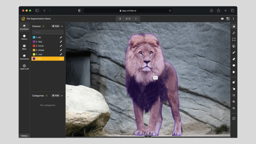
Computer Vision Use Cases | Unitlab Annotate
In this article, we examine the role of computer vision in medical imaging. We will review current applications, explore the benefits for both doctors and patients, and discuss remaining challenges. We will also demonstrate a small-scale project—an X-ray hand segmentation exercise—to illustrate how computer vision can be applied in healthcare.
Computer Vision in Healthcare
Computer vision models, typically developed using deep learning methods and large, diverse datasets, are used to automatically analyze medical scans and help physicians make more accurate decisions. Like other ML models, they require extensive labeled data to detect patterns under various deep learning algorithms. Over time, with a sufficient quantity of AI datasets or ML datasets encompassing a wide range of patient conditions, these models continue to improve.
Consider the case of skin cancer diagnosis: a large annotated dataset with thousands of skin images classified as benign or malignant. AI/ML engineers iteratively train and validate a computer vision model to classify tumors in these two categories.
Over time, given enough varied data, the model can reach an accuracy level comparable to (and sometimes exceeding) that of an experienced physician. Indeed, this is essentially how PathAI (Pathology Transformed) operates in a nutshell. Its automated approach assists doctors in making more informed diagnoses, detecting problems earlier, and ultimately saving lives.
In healthcare, computer vision models have a wide range of applications and bring substantial benefits for both physicians and patients.
Applications of Computer Vision in Healthcare
Healthcare was one of the first industries to adopt computer vision because medical diagnoses heavily rely on imaging—X-ray, CT, and MRI. Although it remains impossible to fully automate diagnostic decisions solely through computer vision, these models serve as supplemental aids for medical professionals, while human expertise remains essential.
Indeed, a hybrid model, blending human insight with computer vision, has been shown to be more accurate than either approach alone.
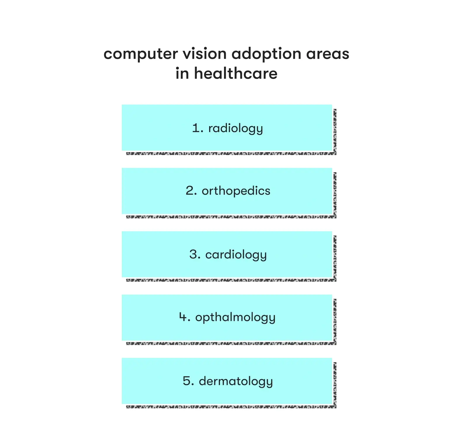
The integration of computer vision into healthcare has produced remarkable progress in multiple medical disciplines:
- Radiology: A specialty that interprets medical images—X-rays, CT scans, and MRIs—to diagnose diseases in humans (and animals). Computer vision models can detect abnormalities, track treatment response, and highlight minute fractures or early-stage tumors that may be missed by manual observation.
- Orthopedics: Concerned with the musculoskeletal system. In orthopedic surgery, computer vision informs preoperative planning and intraoperative navigation by analyzing imaging data to guide precise implant placement and alignment.
- Cardiology: Focused on the heart and cardiovascular system. Computer vision tools assist in diagnosing heart abnormalities, visualizing blood flow during operations, and enabling quantitative assessments of cardiac performance, facilitating enhanced diagnosis and personalized care.
- Ophthalmology: Addresses diseases and treatments of the eye. Here, computer vision is used in the analysis of retina scans, early detection of ocular diseases, and preoperative planning. Eye-tracking technologies also assist in vision correction and tailored therapeutic strategies.
- Dermatology: Concentrates on skin-related conditions. Dermatologists employ computer vision to examine skin lesions and monitor changes over time. High-resolution imaging combined with AI algorithms supports earlier detection of skin cancers and other issues, allowing for prompt intervention.
Benefits of Computer Vision in Healthcare
By adopting computer vision technologies, healthcare providers can realize several advantages for both medical professionals and patients:
- Enhanced Diagnostic Accuracy: AI-assisted image analysis decreases the incidence of diagnostic errors and false positives (suggesting illness where none exists), leading to more precise conclusions.
- Improved Efficiency: Automated review of medical images speeds up the diagnostic process, allowing providers to serve more patients efficiently and reduce costs.
- Early Disease Detection: Computer vision identifies subtle changes in images, promoting the detection of illnesses at an earlier, more treatable stage.
- Personalized Treatment: Detailed image analysis helps tailor treatment approaches to individual patients.
By integrating computer vision models, healthcare systems can not only accelerate the diagnostic process and reduce operating costs but also bolster clinicians in making accurate decisions—essential for patient care. These models aid in reducing both false positives (unnecessary worry) and false negatives (missing a genuine health issue that can be fatal).
Challenges
Like any practical technology, implementing computer vision models in healthcare presents notable challenges in development, testing, and deployment.
1. Data Annotation and Expert Involvement
Medical images are typically more complex, requiring specialized knowledge to identify or outline anatomical structures and pathologies. Thus, image annotation for medical images often calls for the expertise of radiologists or other specialists. Because computer vision models demand highly precise labels, advanced data annotation methods like semantic segmentation are usually required, making the entire process both time-consuming and costly. For medical institutions, this level of data labeling can become a huge burden if not managed carefully. As one radiologist asked:
At what point does starting a medical AI project cross the line into healthcare burden? At what point are you doing harm?
2. Subtle Variations in Medical Imaging
Medical images can show tiny differences between healthy and diseased tissue, making it difficult for algorithms to accurately detect and classify these variations. For instance, a normal chest X-ray can be nearly identical to one indicating early-stage pneumonia, thus requiring models capable of spotting very small indicators within negligible background noise. This is unlike general computer vision tasks (e.g., distinguishing cats from dogs) where the contrast is much greater.
3. Limited and Imbalanced Datasets
It is often impractical to acquire extensive, balanced medical imaging collections due to patient privacy and the rarity of certain pathologies. This shortage limits the development of robust ML models, which typically depend on large, varied AI datasets to generalize effectively. Moreover, class imbalances can lead to biased models that excel at detecting common conditions but fall short in identifying rare disorders.
4. Ethical and Regulatory Considerations
The introduction of AI into healthcare entails navigating a complex ethical and regulatory framework. Protecting patient privacy, obtaining consent for data usage, and adhering to strict legal standards are vital. In addition, there is a moral obligation to ensure that AI systems do not exacerbate existing healthcare inequalities, which calls for careful evaluation of biases in both data and algorithms.
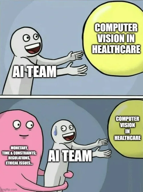
Addressing these issues demands not only technical expertise but also policy-making, financial resources, and broader societal efforts. This mix of challenges helps explain why AI-driven computer vision has yet to achieve wide-scale implementation in healthcare, even though collaborative models (combining clinicians and CV) have been shown to outperform purely human- or machine-based approaches.
Demo Project: X-Ray Hand Semantic Segmentation
Despite extensive investments in healthcare-focused computer vision, data acquisition and labeling remain substantial hurdles. Because medical data must be meticulously annotated, many turn to specialized image labeling service providers. In practice, pixel-perfect segmentation (semantic segmentation) - categorizing every pixel into a class - is often used for its accuracy. However, this approach is markedly more expensive than simpler bounding box methods. Many modern platforms offer auto data annotation features to help reduce costs.
Here, we will demonstrate how to annotate X-ray hand images using semantic segmentation in Unitlab Annotate, an automated data annotation platform. First of all, visit the platform and create a free account.
Project Setup
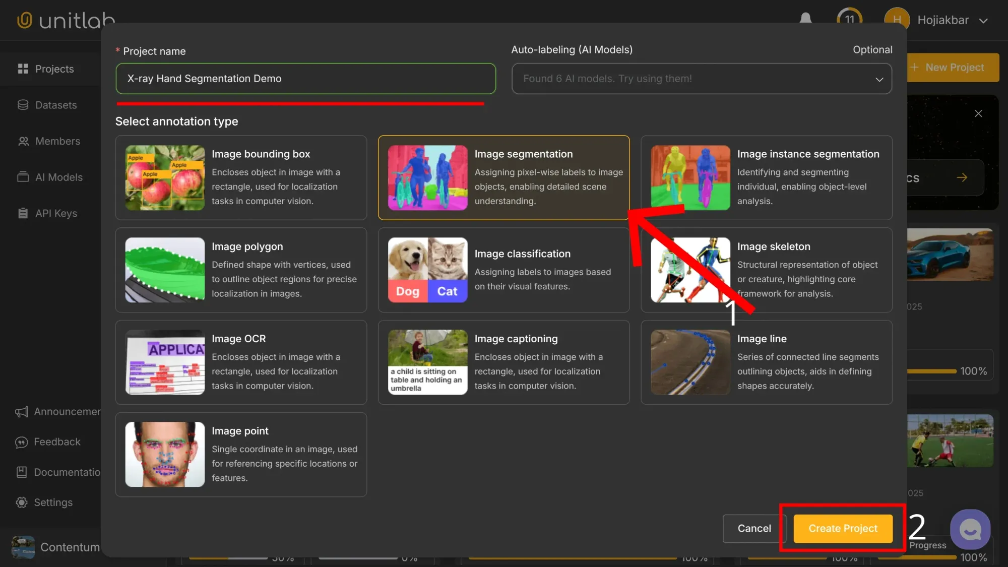
Visit the Unitlab platform, create a project named X-ray Hand Segmentation Demo and select Image Segmentation as the annotation type:
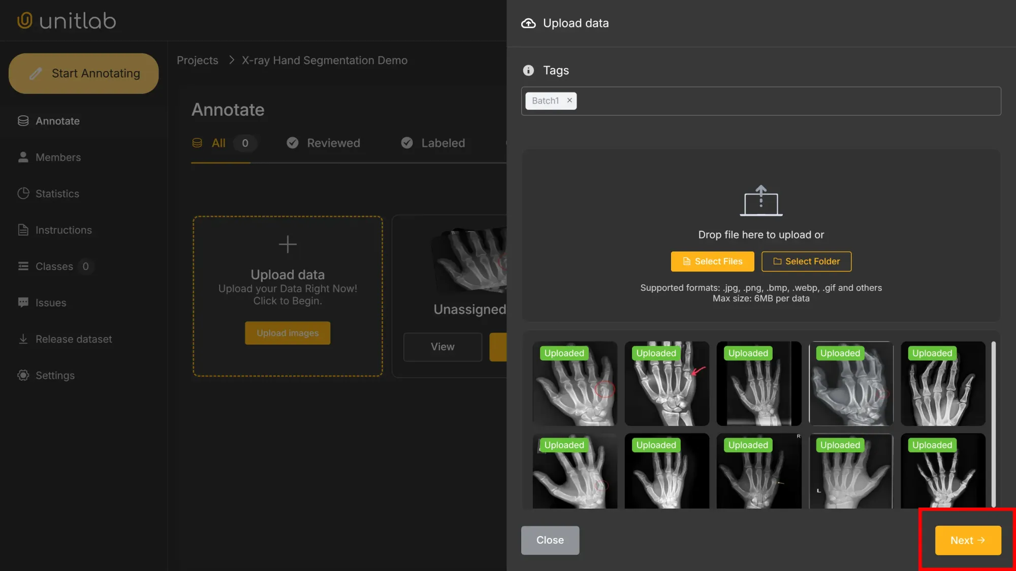
Next, upload the sample X-ray images and assign them to annotators. The images used for this demo are available here.
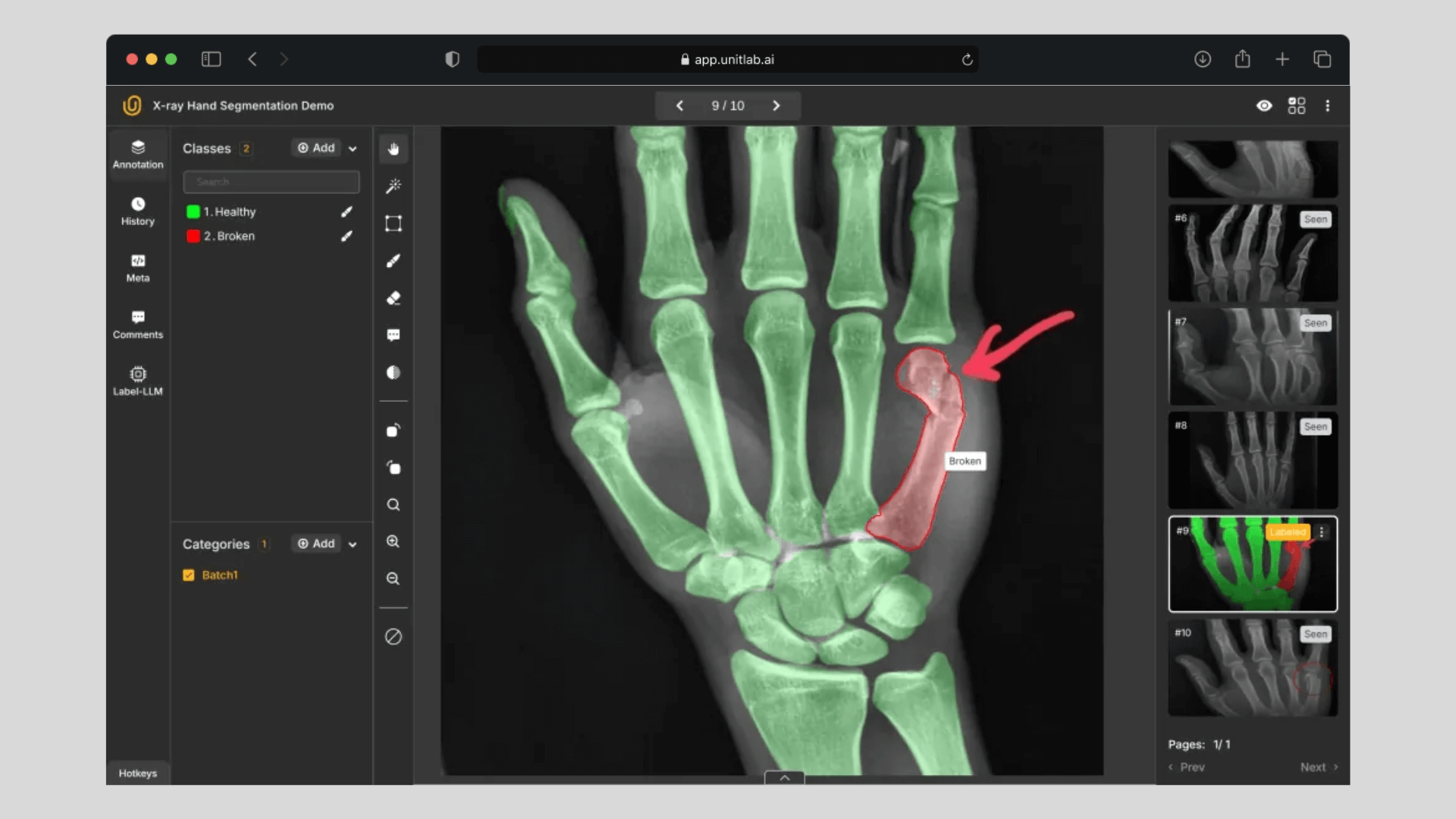
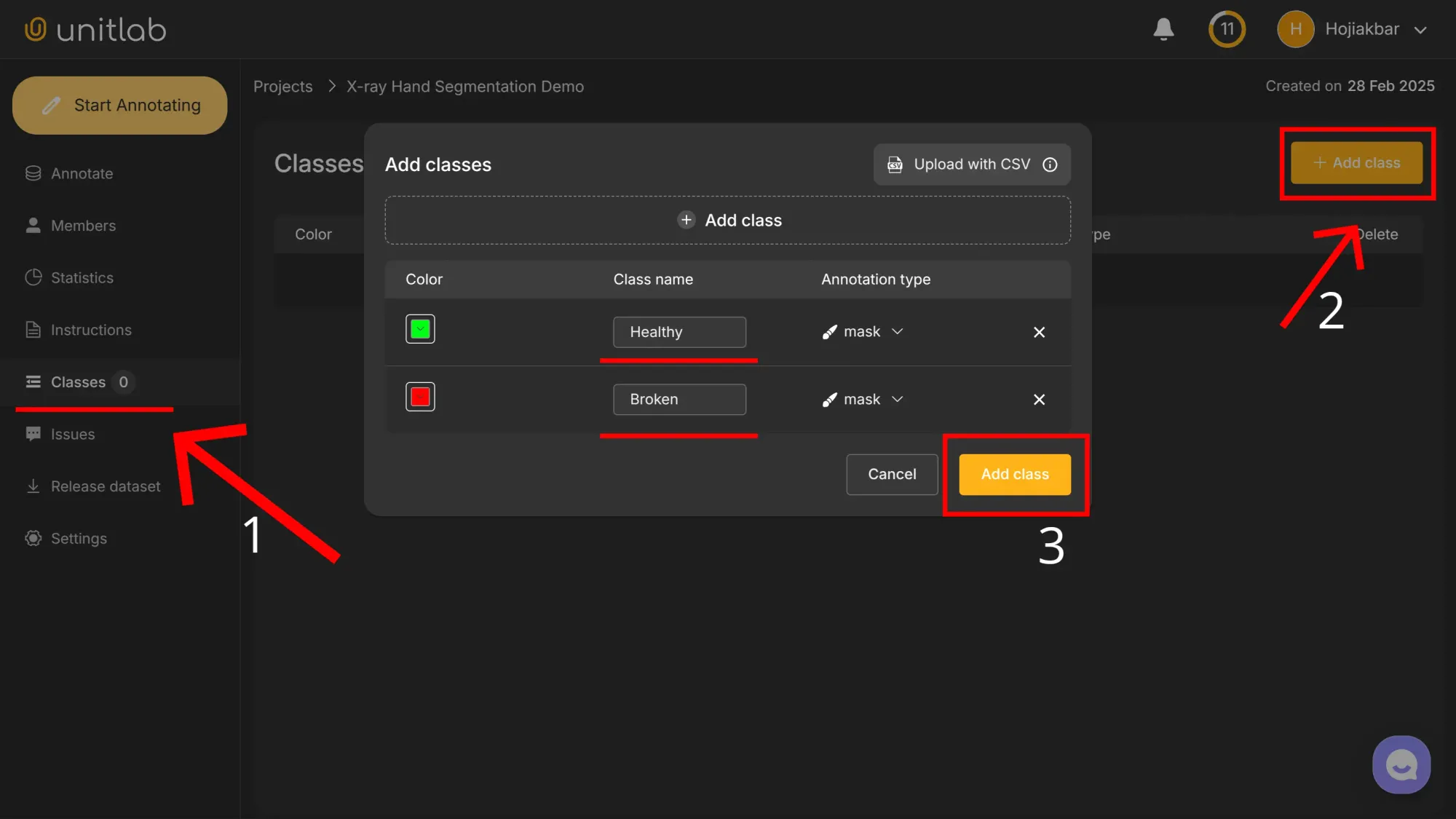
Add two classes, Healthy and Broken, to label the bones appropriately:
Data Labeling
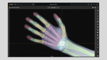
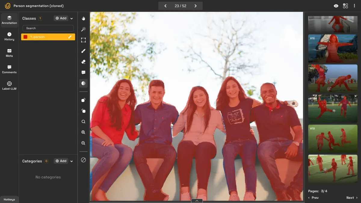
Semantic segmentation involves tagging each pixel with a category. Within Unitlab Annotate, you can apply a brush tool to precisely mark healthy bones, demonstrating a manual image annotation approach. However, it takes a considerable length of time to annotate. The labeling video below originally took 5 minutes:
Manual Semantic Segmentation | Unitlab Annotate
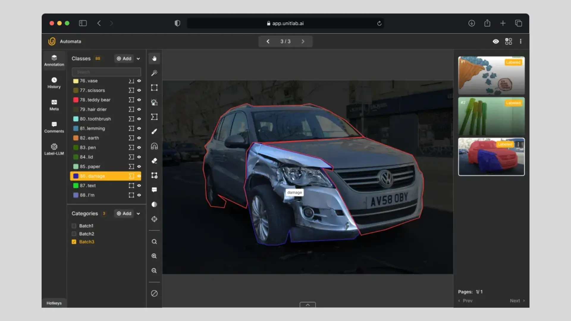
For a faster workflow, the platform’s SAM-powered Magic-Touch can automatically detect image segments and suggest relevant classifications, functioning as an image annotation solution that incorporates auto labeling tools:
SAM-powered Semantic Segmentation | Unitlab Annotate
Dataset Release
You can release a dataset (version 0.1) for training your computer vision model even though data labeling is not complete. Waiting until all images are annotated would beat practicality. Because AI/ML development is iterative, you can release new versions of datasets as you need. In our case, for demonstration purposes, we release an initial dataset even though it contains only two labeled images.
Go to Release Dataset and release a dataset by choosing COCO as the dataset format:

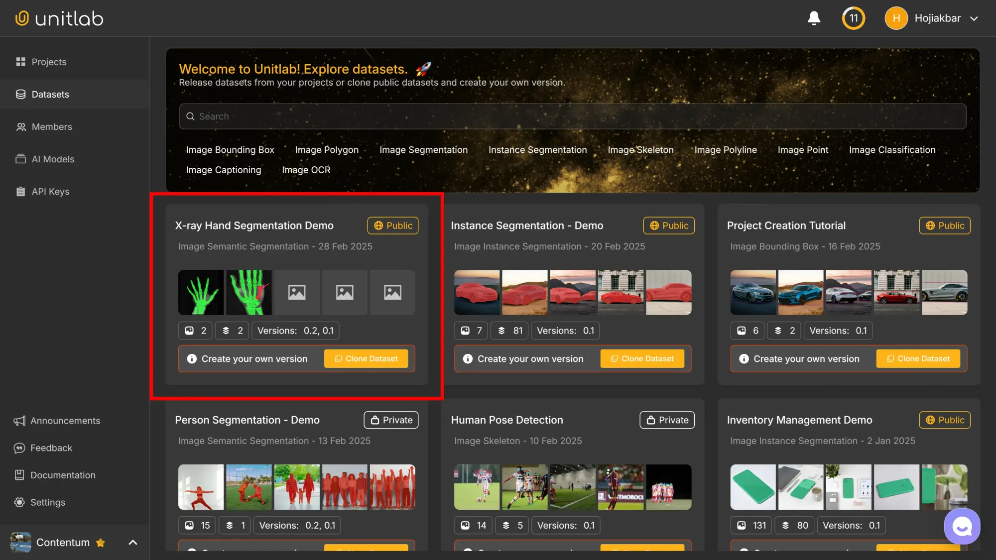
Under the free plan, released datasets become publicly accessible through Unitlab Annotate for usage, download, or cloning:
Refer to this tutorial for more on AI dataset management workflows and best practices in dataset version control:
Dataset Management | Unitlab Annotate
Conclusion
Computer vision has strong potential to revolutionize numerous sectors reliant on visual data, and in some areas it already has. Healthcare, however, stands out for its high level of domain specialization and the limited availability of suitable training datasets.
Despite significant investments of time and capital, along with challenges in data collection and labeling, computer vision offers valuable benefits to doctors, patients, and hospitals—particularly in fields like radiology.
To illustrate the intricacies of data annotation service in healthcare, we created and labeled a sample X-ray hand dataset using semantic segmentation in Unitlab Annotate. While this was merely a small-scale demonstration, large projects follow similar fundamentals, albeit with greater complexity. We invite you to explore these methods and tools—to effectively manage ML datasets.
Explore More
For further reading on practical applications of computer vision, check out these resources:
- Practical Computer Vision: Parking Lot Monitoring
- The Transformative Power of Computer Vision in Logistics
- Enhancing Inventory Management with Computer Vision
References
- Darvin Yi. (No date). The Unique Problems of Medical Computer Vision. Sirona Medical: Source
- Darya Efimova. (Jun 18, 2024). Computer Vision in Healthcare: a Secret Guide for Winners. EPAM Startups: Source
- Dora Mekouar (Feb 19, 2025). Does AI detect breast cancer better than doctors can? Voice of America News: Source
- Krista Conger. (Apr 11, 2024). AI improves accuracy of skin cancer diagnoses in Stanford Medicine-led study. Stanford Medicine: Source